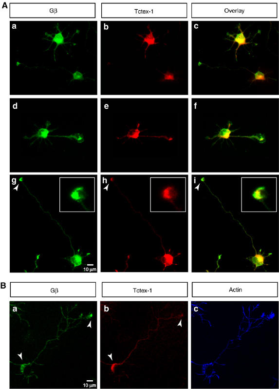
Figure 5.

Overlapping distribution of Tctex-1 and Gβ in hippocampal neurons. (A) Cultured hippocampal neurons in various stages of differentiation were colabeled for Gβ (green in a, d and g) and Tctex-1 (red in b, e and h). The overlayed images are shown in c, f and i. Gβ and Tctex-1 show homogenous expression within the cell body and all the neurites in typical stage 2 cells (a–c). As the neurons progress through stage 3, Gβ and Tctex-1 continue to overlap within the cell body, but in addition, show strong co-distribution at the growth cones of the future axon (a–i). Majority of the stage 3 neurons examined (70%, n=100) (a–f) show enhanced colabeling of Gβ and Tctex-1 in axonal growth cones as compared with the minor neurites that do not show an enrichment of Gβ or Tctex-1 at the tips. A small subset of the stage 3 neurons examined (30%, n=100) continued to show colabeling of Gβ and Tctex-1 in the cell body and at the axonal growth cones, and also showed some colabeling at the tips of the minor neurites (g–i). A magnified view of the growth cone of a stage 3 neuron is shown in the inset (g–i) Greater than 100 individual neurons were examined. Scale bar equals 10 μm (a–i) and 3 μm in the magnified insets (g–i). (B) Confocal image of a representative stage 3 neuron shows perinuclear, cytoplasmic staining for Gβ and Tctex-1 within the cell body and an enrichment at the tips of some of the axonal growth cones (panels A–C). Scale bar equals 10 μm.
