
Figure 1.
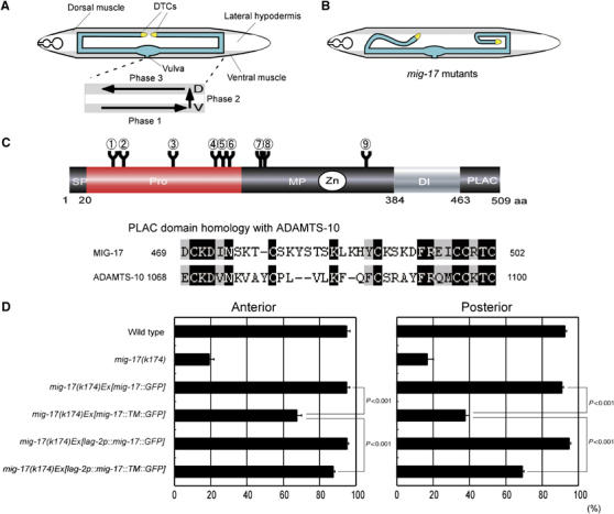
Tissue-specific expression of a membrane-anchored MIG-17. (A) The gonad morphology and the phases of DTC migration in wild-type hermaphrodites. (Upper) The gonad is shown in blue. Ventral and dorsal body wall muscles are shaded. Unshaded part between dorsal and ventral muscles corresponds to the hypodermis. (Lower) The phases of DTC migration are shown by arrows. (B) Abnormal gonad morphogenesis in mig-17 mutants. (C) (Upper) Domain structure and potential N-glycosylation sites of MIG-17. The circled numbers (1–9) indicate potential N-glycosylation sites (N-X-S/T). 1, N52; 2, N65; 3, N123; 4, N172; 5, N183; 6, N189; 7, N218; 8, N219; 9, N350. (Lower) Sequence alignment of the PLAC domains of MIG-17 and human ADAMTS-10. Identical and homologous amino acids are shown in black and gray, respectively. (D) A membrane-anchored MIG-17 can rescue mig-17 mutants when expressed in DTCs. The ratios of normal DTC migration are shown in bar graphs with the mean±s.e.m. For the animals carrying lag-2 promoter-driven transgenes, those with GFP expression in DTCs were scored. P-values from Fisher's exact test are shown for some combinations. n=120. DI, disintegrin domain; PLAC, protease and lacunin domain; MP, metalloprotease domain; Pro, prodomain; SP, signal peptide.