
Figure 5.
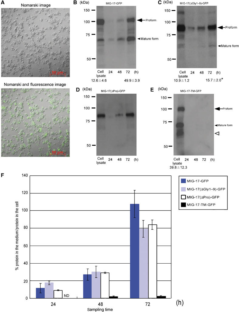
Secretion of MIG-17 from primary culture cells. (A) A typical culture of C. elegans embryonic cells 4 days after plating. Nomarski (upper) and combined Nomarski and fluorescence (lower) images of MIG-17-GFP-expressing primary culture cells. (B–E) Secretion of MIG-17-GFP, MIG-17(ΔGly1–9)-GFP, MIG-17(ΔPro)-GFP and MIG-17-TM-GFP into the media. The culture media were sampled at the indicated time points and cell lysates were prepared from 72-h cultures. The samples were immunoblotted with anti-GFP. The same set of experiments (from culture to immunoblot) was independently performed twice. The ratios of mature form to the sum of pro- and mature forms are shown for some lanes as the mean±s.e.m. The asterisk indicates that the rate of conversion from the proform to mature form was significantly slower in MIG-17(ΔGly1–9)-GFP-expressing cells compared with MIG-17-GFP-expressing cells (Student's t-test; P<0.05). The smeared band migrating slightly faster than the mature form (open arrowhead) in (E) appears to be partially degraded proteins. (F) Kinetics of secretion of MIG-17-GFP fusion proteins. The ratios of secreted MIG-17-GFP fusion protein to MIG-17-GFP fusion protein retained in the cell lysates were plotted against sampling times. For MIG-17-GFP, MIG-17(ΔGly1–9)-GFP and MIG-17-TM-GFP, the intensitiesy of the bands for the pro- and mature forms were summed and used for calculation. The error bars represent the mean±s.e.m.