
Figure 6.
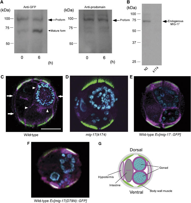
MIG-17 is secreted and localizes to the gonad as a proform. (A) The antibody against the MIG-17 prodomain recognizes the MIG-17 proform but not the mature form. Extracts from wild-type worms expressing MIG-17-GFP were incubated for 0 and 6 h at room temperature and analyzed. The proform and the mature form were recognized by anti-GFP (left panel), but only the proform was detected by anti-MIG-17 prodomain (right panel). The faint bands in the right panel seem to be nonspecific binding of anti-prodomain because these also appeared in the immunoblot of the mig-17 mutant extract after long exposure. (B) Detection of endogenous MIG-17. About 200 μg of TCA-precipitated lysate from wild-type or mig-17 mutant animals was immunoblotted with the anti-MIG-17 prodomain. (C–F) Immunohistochemistry using anti-MIG-17 prodomain (C, D) and anti-GFP (E, F). Cross-sections of hermaphrodites for wild-type (C), mig-17 mutant (D) and wild-type carrying mig-17∷GFP (E) or mig-17(D79N)∷GFP (F) were stained. Merged images of the specimens stained with antibodies (pink), fluorescein–phalloidin (green) and DAPI (blue) are shown (dorsal to the top). Fluorescein–phalloidin stains actin filaments in dorsal and ventral body wall muscles. Arrowheads indicate prodomain localization in the gonadal basement, whereas arrows indicate localization in the hypodermal basement. Scale bar, 20 μm. (G) Schematic presentation of a wild-type cross-section depicting anti-prodomain signals shown in pink.