
Figure 2.
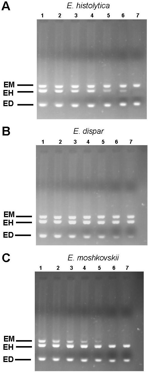
Detection of E. histolytica (A), E. dispar (B) and E. moshkovskii (C) in mixed cell lysates. To 1000 cells of E. dispar and E. moshkovskii (A) or E. histolytica and E. moshkovskii or (B), E. histolytica and E. dispar (C), 1000 cells (lane 1), 100 cells (lane 2), 10 cell (lane 3), 1 cell (lane 4), 0.1 cell (lane 5), 0.01 cell (lane 6) and 0.001 cell (lane 7) of the other species were added. Amplification was done by nested multiplex PCR.