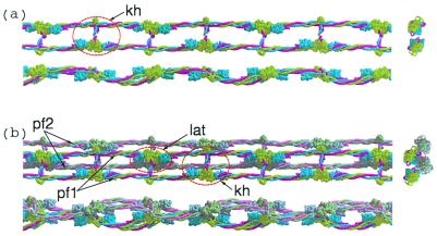
Figure 5.
(a) Three views (top, side, and end-on) of a single protofibril formed by knob-hole (kh) interactions. Light green, γ-chains; blue, β-chains, red, α-chains. End-on view is shown at the right side. (b) Lateral association (lat) of two protofibrils (PF1 and PF2, top and side views; end-on view is shown at right).

