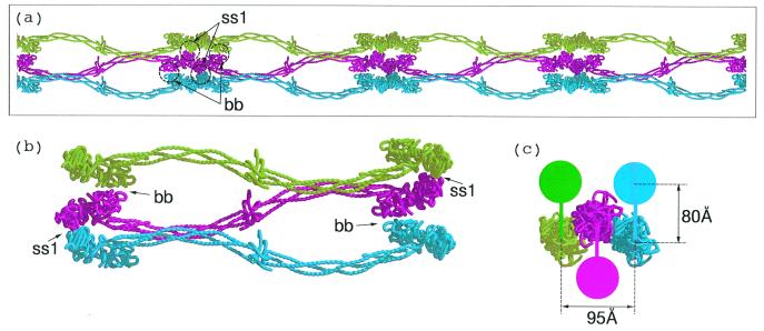
Figure 6.
(a) Three protofibrils associated by lateral involvement of γC domains; only one of the strands from each protofibril is shown to facilitate visualization. In order for a fiber to grow the lateral interactions involving different halves of a unit must extend to different protofibrils. (b) The γC associations (denoted ss1) are depicted as blue to red and red to green. The βC associations (denoted bb) follow the same pattern except red to green and blue to red. The hypothetical interaction between βC domains uses regions that are homologous to γ-γ end-to-end faces. (c) End-on view of three associated protofibrils. The solid colored discs denote the molecular units of the protofibril not included in a and b; the colored vertical bars denote knob-hole interactions. The 80 Å-distance is taken from Fig. 3a; the 95 Å-distance between centers of associated protofibrils is taken directly from the lateral packing distances observed in DD-BO.

