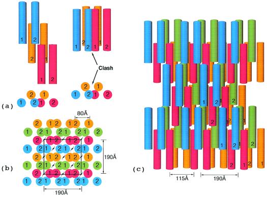
Figure 7.
(a) End view of simplest highly ordered model fibrin fiber with xy face of hypothetical unit cell outlined. The unit cell dimensions are 190 × 190 × 460 Å. The first 190 Å dimension is based on distances observed in the crystal packing in DD-BO (Table 1). The second 190-Å distance is a consequence of the two strands of any protofibril being 80 Å apart. A unit cell with the same dimensions has been reported for a neutron diffraction experiment with fibrin oriented in a magnetic field (38). (b) Top view of fiber showing 115 Å-wide cavities resulting from half-staggered overlaps; the 115-Å dimension is calculated directly from unit cell.

