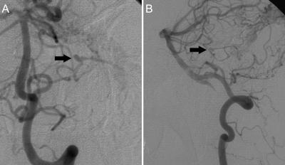
Figure 2.
(A) Anteroposterior and (B) lateral view follow-up angiograms, left vertebral artery injection, after the first-stage operation show the 3-mm left AICA feeding-artery aneurysm beyond the floccular segment (arrows) that developed during the interval. AICA, anterior-inferior cerebellar artery.

