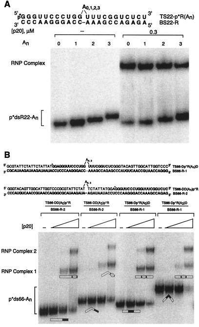
Figure 2.
Mobility-shift assays of An-bulged duplexes. RNA is bold, DNA is in normal lettering, and bulges are indicated. p*dsR22-An and p*ds66-An represent 5′-labeled top strand annealed to bottom strand. (A) Electrophoresis was on a native 10% polyacrylamide gel, with 0 or 0.3 μM p20. Gel was run at 22°C, resulting primarily in the 1:1 protein-RNA complex at this [p20]. (B) Electrophoresis was on a native 15% polyacrylamide gel with 0, 0.01, 0.03, and 0.1 μM p20. Gel was run at 4°C. Positions of 1:1 and 2:1 RNA-protein (RNP) complexes are shown. Duplex structures in the 1:1 complex are drawn on the basis of the best model. Straightening may not be complete. Protein is depicted as an oval and the duplex as a rectangle with dsRNA segments filled and RNA–DNA hybrid segments open.

