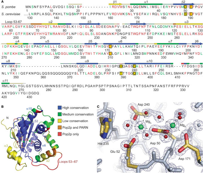
Figure 1.
Overview of the S. pombe Pop2p structure. (A) Alignment of Pop2p from S. pombe and S. cerevisiae. Fully conserved residues are shown in red and conserved functionality in green. The secondary structure of S. pombe Pop2p is indicated above the sequence and colour-coded according to similarity to other homologous structures identified by a DALI search (see text) with the extremes being structural elements solely found in Pop2p (red) and regions found in most or all of the homologous structures (blue). Other colours represent intermediate occurrence as indicated in (B). The five active site residues are shown framed on a yellow background. Sequence diagram was produced with SecSeq (39). (B) Cartoon representation of S. pombe Pop2p. The structure is colour coded as indicated and described in (A). The side chains of the active site residues are shown as sticks and coloured by element, whereas ions in the active site are shown as green spheres. All structure figures were produced with PyMOL (40). (C) Close-up of the active site. Active site residues are coloured by element and associated magnesium ions are shown in green, waters involved in coordination of the active site ions are coloured red, and the octahedral coordination of the active site magnesium ions is indicated by dashed lines. The refined 2mFo-DFc electron-density map is shown contoured at 2.5σ.

