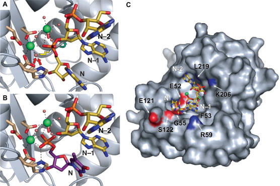
Figure 3.
Pop2p–RNA interactions. (A) The pre-hydrolysis state as illustrated by superposition of poly-A from the structure of human PARN (PDB entry 2A1R, yellow sticks) onto the S. pombe Pop2p structure. The 3′ nucleotide (N), the penultimate nucleotide (N–1) and the third last nucleotide (N–2) are shown. Arrows indicate the hydrolytic reaction mechanism in which a water molecule associated with one of the active site ions is activated (red arrows) for attack on the most 3′ phosphate of the RNA (yellow lightening) leading to breakage (green arrow) of the bond between the phosphate group and the N–1 nucleotide. (B) The post-hydrolysis state illustrated by superposition of dTMP from the ε-subunit of E. coli DNA pol III (PDB entry 1J54, purple sticks) showing the position of the 3′ nucleotide (N) immediately after cleavage. The N–1 and N–2 nucleotides (PDB entry 2A1R, yellow sticks) from the PARN poly-A illustrate the position of the new 3′ end of the substrate. (C) Possible Pop2p–RNA contacts. Poly-A from PARN (PDB entry 2A1R, yellow sticks) superposed on the active site region of the S. pombe Pop2p, shown as a surface representation with atoms of residues possibly involved in the RNA recognition coloured by element.

