Figure 5.
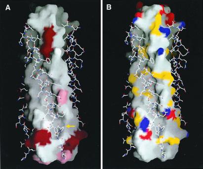
The surface properties of the HRSV-N57 trimer with the HRSV-C45 peptides displayed in a stick-style representation. View same as Fig. 4C. (A) Surface variability of the HRSV-N57 trimer (analysis based on RSV sequences available in GenBank and Swiss-Prot). The residues shown in dark red vary among different human virus strains. The residues shown in pink are identical among 20 human strains of HRSV but are different in bovine respiratory syncytial virus. The cavity region is indicated by a yellow arrow. (B) Surface mapping of groups with the potential to form electrostatic and polar interactions. Nitrogen and oxygen atoms from charged amino acid side chains are shown in blue and red, respectively. Nitrogen and oxygen atoms from polar amino acid side chains are shown in yellow. Figure was drawn with the program grasp (46).

