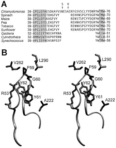
Figure 3.
The Rubisco small subunits of plants and green algae contain a long loop between β strands A and B. (A) Sequences of the βA/βB region of small subunits from various species (5, 14, 21, 37–39). The N54S and A57V suppressor substitutions in the small subunit of Chlamydomonas are denoted with arrows. Locations of β strands A and B (gray highlighting) are assigned from the x-ray crystal structures of spinach (21), tobacco (40), Galdieria partita (38), and Synechococcus (41) Rubisco. (B) Stereo view of large- and small-subunit structural features within the x-ray crystal structure of spinach Rubisco (21). Leu290 and Val262 reside in one large subunit, whereas Ala222 is in a neighboring large subunit. These residues are identical to those in Chlamydomonas Rubisco. An L290F substitution in the large subunit of Chlamydomonas is complemented by A222T and V262L large-subunit substitutions (7, 13, 20). Arg53, Pro59, Gly60, Tyr61, and Tyr62 (Arg59, Cys65, Leu66, Tyr67, and Tyr68 in Chlamydomonas Rubisco) reside within the small-subunit βA/βB loop, which is represented as a ribbon. An R53E substitution in the small subunit of pea Rubisco blocks holoenzyme assembly (43).

