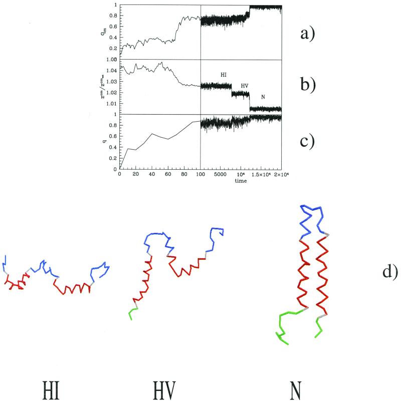
Figure 3.
Typical time dependence of different parameters as a function of the Monte Carlo steps for the pathway U → HI → HV → N. Fraction of native contacts inside the membrane (a), normalized z coordinate of the center of mass of the protein (with respect to that of the native state conformation) (b) and overall fraction of native helical contacts (c). Each Monte Carlo step corresponds to 50,000 attempted local deformations. The transition from state HI to state HV is signaled by a sharp jump of the position of the center of mass. Note that there is no perceptible sign of this transition in terms of newly formed native contacts. Most of the helical contacts are formed in the early stages of the folding. This fraction does not significantly increase until helices correctly assemble and the interhelical contacts are formed. The HV → N transition is reached by a progressive zippering of the horizontal and vertical helices. This zippering is usually very quick (few Monte Carlo steps) and is only slightly slowed down (see the plateau corresponding to qm ≈ 0.9 in a) when the trajectory passes through somewhat deformed conformations. (d) Protein conformations at different times during the folding. The colors red, green, and blue have the same significance as in Fig. 1a with the grey bonds being ones crossing the membrane.

