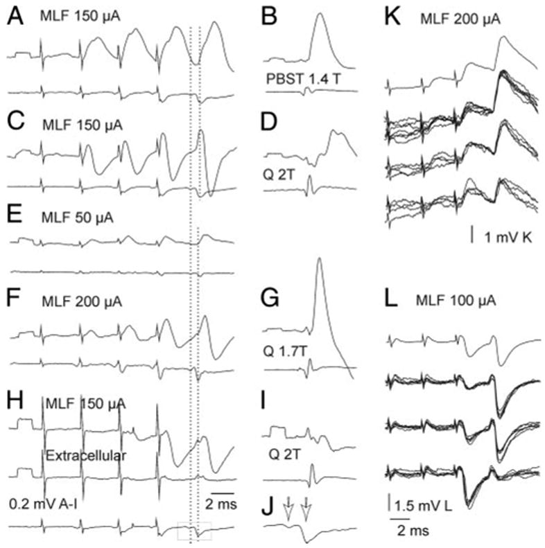
FIG. 3.

EPSPs and IPSPs evoked from MLF in Ib interneurons. A–B, C–D, E–G, and H–I: intracellular averaged records from 4 Ib interneurons projecting to motor nuclei (top traces), records from the cord dorsum (bottom traces) and extracellular records from outside the last interneuron (middle trace in H). A and C: monosynaptic EPSPs of MLF origin, followed by disynaptic EPSPs (arrows in A) or IPSPs. E and F: EPSPs classified as evoked disynaptically, followed by IPSPs at higher stimulus intensities. H: disynaptic IPSPs. J: twice expanded MLF volleys boxed in H; arrows indicate the direct and relayed components. Dashed lines indicate positive peaks of the 2 components. EPSPs with the onset between these peaks were considered as evoked monosynaptically. K and L: averaged (top) and single records from 2 additional interneurons. Those of a similar size are superimposed separately.
