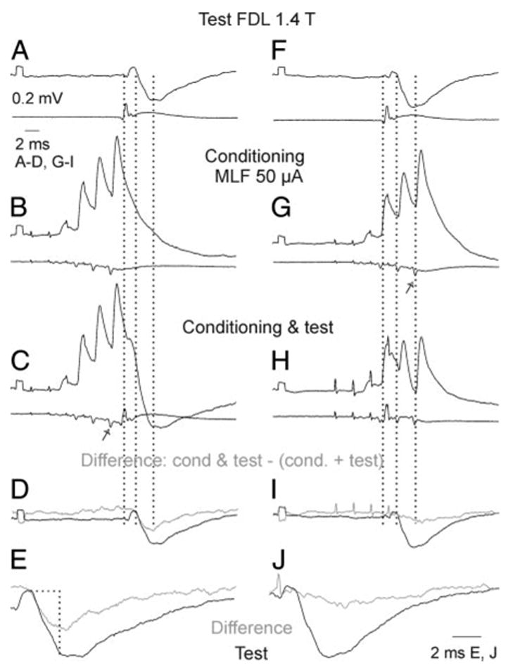
FIG. 7.

Facilitation of Ib IPSPs recorded in a GS motoneuron by conditioning stimulation of the contralateral MLF. Pairs of records in A–C and F–H are from a GS motoneuron and from the cord dorsum. A and F: PSPs evoked by test stimuli alone. B and G: PSPs evoked by conditioning stimuli alone. C and H: PSPs after both these stimuli. D–J: differences between the conditioned and test IPSPs (gray) superimposed on the test IPSPs (black), at the same scale and twice expanded. Areas that were measured are enclosed by the horizontal and vertical lines in E. Test and conditioning stimuli were timed in such a way that group I volleys indicated by the first dotted line in A–C and F–H were preceded by the last relayed MLF volley (arrow in C) in left panels or were preceded by the second MLF volley in right panels. Consequently the Ib IPSPs were superimposed on the decay phase of EPSPs evoked from MLF in C and coincided with these EPSPs in H. Second and third dotted lines indicate onset of the IPSPs and the end of the time windows used for the measurements of the areas of the IPSPs. Calibrations in A are for all records except E and J. Note that the relayed volleys reflecting activation of commissural interneurons were marginal after the first stimulus and increased after the successive stimuli. All records are averages of 20 single records.
