Figure 7.
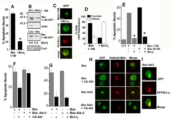
The Bax N-terminus regulates susceptibility to inhibition by anti-apoptotic molecules. A, Apoptotic nuclear damage (15 hours post-transfection) was assessed in HEK cells transfected with Bax 1–146-GFP with or without RFP-Bcl-xL. B, HEK cells transfected with Bax 1–146 GFP and Bcl-xL were harvested after 15 hours of culture and Bcl-xL immunoprecipitated from the cell lysate. The precipitated complex was analyzed for Bax 1–146-GFP and actin by western blot analysis. WCL is the CHAPS buffer-solubilized whole cell lysate used in the immunoprecipitation. HEK cells transfected with Bax 1–146-GFP were harvested 15 hours post-transfection and processed to enrich for the cytosol (Cyto) and the membrane/mitochondrial (Mito) fractions. The distribution of GFP and Cox4 was determined by western blot analysis. WCL represents the whole cell lysate. Numbers below the blots are the densitometric analysis of the GFP signal in the specific fraction relative to the total in the mitochondria and cytosol fractions. C, HEK cells expressing Bax 1–146 alone or Bax 1–146+RFP-BclxL were assessed for distribution as described in 6B. E, Apoptotic nuclear damage was assessed in HEK cells 18–20 hours following transfection with combinations of constructs shown in the panels. F and G, Regulation of Bax and Bax-Ala3 induced apoptosis by constitutively active (CA)- Akt (F) and Bcl-xL (G). The data in all panels are presented as mean ± SD and are derived from three-five independent experiments. H and I, HEK cells transfected with Bax-GFP or Bax-Ala-3 with CA-Akt and Mito-dsRed2 or RFP-Bcl-xL where indicated were harvested 15 hours post-transfection. Confocal images for distribution of GFP tagged constructs of Bax (green) and Mito-dsRed2 (red) or RFP-Bcl-xL (red) were assessed for co-localization (Merge). Scale Bar: 10 microns. Representative images (each corresponding to a single image field) are shown. *p < 0.001

