
Figure 3.
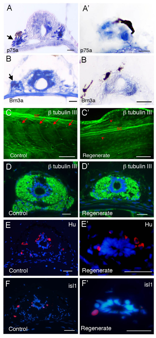
Expression of neural markers in spinal cord of normal and regenerating tails. (A-B') In situ hybridization detection of mRNA expression of p75a (A, A') and Brn3a (B, B') on transverse sections. (A-B) Control tails. (A'-B') Tail regenerates. Arrows in (A, B) indicate ganglia and the arrow in (A') indicates a sporadic ganglion cell. (C-D') β III tubulin expression (green) in whole mount tadpole tails (C, C') and on cross sections (D, D'). (C', D') are one-month regenerates. (E-F') Expression of Hu (E, E') and islet 1 (F, F') are detected by antibody staining on transverse sections. (E, F) un-operated control, (E', F') 2 week old tail regenerates. Scale bars: 250 μm in (E, E'), 20 μm in the rest.