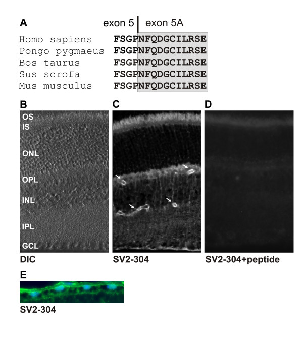
Figure 5.

Localization of the ABCC5_SV2 protein in mouse retina. (A) Alignment of the immediate C-termini of putative ABCC5_SV2 proteins from several mammalian species reveals 100% amino acid identity. The relative location of the exon 5 – exon 5A boundary is shown by a vertical bar. (B) Differential interference contrast (DIC) image shows the various retinal cell layers: OS, outer segments; IS, inner segments; ONL, outer nuclear layer; OPL, outer plexiform layer; INL, inner nuclear layer; IPL, inner plexiform layer; GCL; ganglion cell layer. (C) ABCC5_SV2 immunolabeling of frozen mouse retina sections with the SV2-304 antibody. Arrows point to labelled endothelial cells of the inner retinal blood vessels. (D) Mouse retinal section stained with SV2-304 antibodies preincubated with an excess of competing GST-ABCC5_SV2 peptide. (E) RPE monolayer labelled with SV2-304 antibodies (green), the nuclei were counterstained with DAPI (blue). The basal side of the RPE is oriented towards the top.
