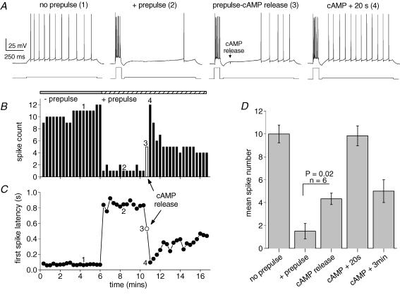
Figure 6. The kinetics of cellular excitability changes following rapid release of cAMP.
A, the four panels show, from left to right, spike pattern during mild depolarization from a membrane potential of −63 mV (no prepulse); inhibition of firing by sAHP (+ prepulse); relief of inhibition immediately following cAMP release (prepulse-cAMP release); complete block of inhibition 20 s after cAMP release (cAMP + 20 s). The numbers for each trace refer to the time points indicated in B and C. B, spike count during the 1 s depolarizations of the experiment shown in A. Addition of the prepulse to evoke a slow AHP dramatically reduced spike numbers. The open bar denotes the point of photorelease of cAMP. The return to control excitability (4) representing complete suppression of the slow AHP is short-lasting. C, first spike latency during the 1 s depolarizations of the experiment in A. The slow AHP retards spiking until the release of cAMP (open symbol). D, pooled data (mean ± s.e.m.) for 6 experiments using the protocol in A. Mild depolarization evoked 10 ± 0.8 spikes which was reduced to 1.5 ± 0.7 spikes in the presence of a prepulse to generate sAHP. Significantly increased spiking (4.3 ± 0.5) was observed immediately after cAMP release and after 20 s excitability was indistinguishable from control activity (9.8 ± 0.9 spikes). Partial relief of cAMP action was seen after 3 min (5 ± 1 spike).

