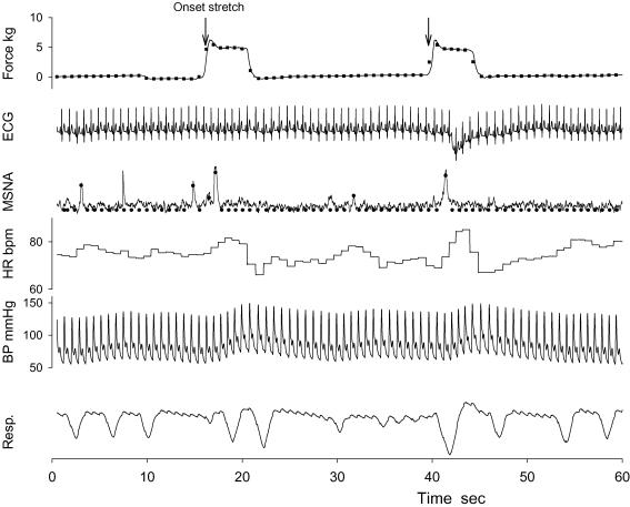
Figure 1. Representative tracing of stretch force, ECG, muscle sympathetic nerve activity (MSNA), heart rate (HR), arterial blood pressure (BP), and respiratory excursion (Resp.) during two passive stretch bouts.
The stretch bouts were repeated 25 times in each trial, and the intervals between 2 bouts were of random length (15–25 s). The filled circles and squares represent the processed beat-by-beat data of MSNA and force, respectively. The arrows indicate the beat of the onset stretch, and were used to align the data segments of the beat-by-beat data in signal averaging.

