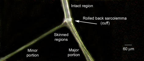
Figure 1. Skinning a skeletal muscle fibre by microdissection.
A single skeletal muscle fibre was dissected away from a rat extensor digitorum longus muscle under paraffin oil. Individual myofibrils within the fibre run axially, and a group of myofibrils (‘minor portion’) were pulled away from the fibre, causing the entire sarcolemma along the fibre segment to roll back as a ‘cuff’ of membrane, which then could be dissected away and analysed separately from the resulting ‘skinned’ fibre portions that remain (major and minor portions pooled together as required).

