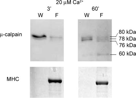
Figure 4. Cytosolic and bound pools of μ-calpain are both autolysed at elevated [Ca2+].
Western blot showing that μ-calpain residing in both the cytosolic and bound pools in quiescent fibres is autolysed in a Ca2+- and time-dependent manner. Skinned extensor digitorum longus fibre segments (examined in pairs for better detection) were washed for 60 s in 20 nm Ca2+ solution to separate cytosolic pool of μ-calpain, and then cytosolic constituents (W) and fibre segments (F) were separately exposed for 3 or 60 min to a solution at 20 μm free [Ca2+].

