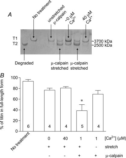
Figure 6. Proteolysis of titin by activated exogenous μ-calpain.
A, 2.8% SDS-PAGE gel showing silver staining of titin in rat extensor digitorum longus fibres following passive force measurements and treatments as in Figure 5. Fibres with ‘no treatment’ were skinned but not mounted on a transducer. Degraded titin was produced by incubating fibres at 30°C for 60 min. B, mean percentage (±s.e.m.) of total titin in full-length form in fibres given indicated treatments. One of the stretched fibres exposed to μ-calpain broke during re-stretch and could not be included in the results in Figure 5C. *Significant difference in proportion of full-length titin remaining compared with other treatments (one way ANOVA with Newman–Keuls post hoc test, P < 0.05).

