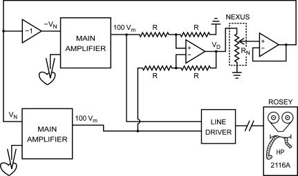
Figure 1. Block diagram of first dynamic clamp setup.
Setup used to study the mutual synchronization of two small clusters (∼100 μm in diameter) of spontaneously active embryonic chick ventricular cells (Scott, 1979). An analog circuit is used to generate positive and negative command potentials proportional to the difference in membrane potential between the two clusters. These command potentials (VN and −VN) are sent to the main amplifiers to let them inject a ‘nexus current’ into each of the clusters such that these are effectively coupled by a nexus resistance RN. The setup includes a ‘minicomputer’ (HP 2116A), but its role is limited to recording the membrane potential of each cluster. Redrawn from Fig 2–6 of Scott (1979).

