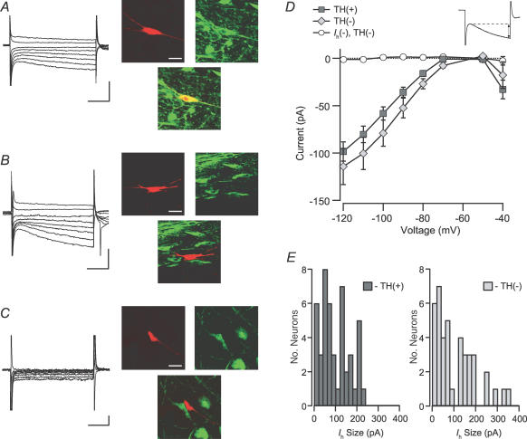
Figure 5. Ih is similar between dopaminergic and non-dopaminergic neurons.
A, example Ih(+) neuron filled with biocytin (red; scale bar: 40 μm) during recording and immunocytochemically identified to be TH(+) (green, yellow; scale bars: 200 pA and 50 ms). B, example Ih(+) neuron that was TH(−) (scale bars: 200 pA and 50 ms). C, example Ih(−) neuron that was TH(−) (scale bars: 20 pA and 50 ms). D, the current induced by stepping the cells in voltage clamp to a variety of membrane potentials was not different between TH(+) and TH(−) Ih(+) neurons. Inset, the Ih was measured as the difference between the initial response to the step and the end of each 200 ms pulse. E, the magnitude of the Ih due to a step from −60 to −120 mV was not different between TH(+) and TH(−) Ih(+) neurons (n = 46 and n = 41, respectively). Ih(−) neurons were excluded from this analysis.

