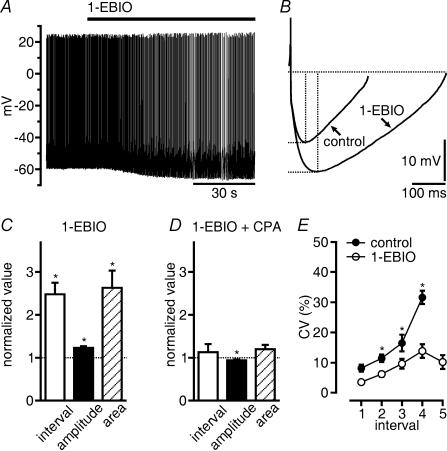
Figure 4. 1-EBIO reduces discharge frequency and increases AHP area.
A, spontaneous firing of a GABA neuron is slowed down by 1-EBIO (200 μm). B, five AHPs in control and in 1-EBIO of the neuron shown in A were averaged and superimposed to illustrate the increase in amplitude and duration. C, summary of the increase in interspike interval, of the amplitude and area of AHPs of 21 GABAergic SNr neurons in 1-EBIO. Mean values of interspike intervals were calculated from intervals between spikes occurring during 90 s and normalized. Amplitude and duration of AHPs were measured as indicated by the dashed lines in B, and averages of 25 events were normalized. 1-EBIO potentiated AHP area more strongly than AHP amplitude. D, CPA (10 μm) applied in 1-EBIO slightly decreased AHP amplitude. CPA had no effect on interspike intervals of spontaneous discharge and AHP area (n = 13 GABAergic SNr neurons, display as in C). E, 1-EBIO increased the regularity of discharge of GABAergic SNr neurons at low spike frequency. Coefficients of variation (CV) were plotted against interspike intervals collected from 21 cells in which frequency of continuous discharge was changed by constant current injection. Each point represents an average of data obtained from 3 to 19 measurements falling into a bin of 200 ms. Continuous discharge only was considered for these neurons. Because this discharge pattern was preserved in 1-EBIO at lower frequencies than in control, not all intervals had a matching control. Numbers on the abscissa denote consecutive intervals of 200 ms in duration beginning at 300 ms. *P < 0.05.

