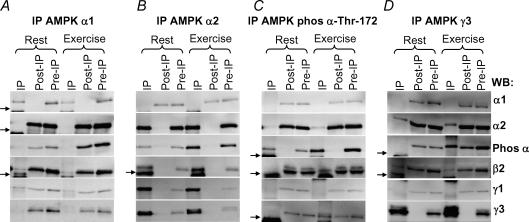
Figure 1. AMPK heterotrimer composition and phosphorylation in human skeletal muscle.
Lysates were prepared from biopsies taken in the vastus lateralis muscle before and after 20 min of exercise at 80%V˙O2,peak (n = 11). From 400 μg of lysate AMPK α1 (A), α2 (B), phospho α-Thr-172 (C) or γ3 (D) was immunoprecipitated (ip). The figure shows representative blots of the IP, post-IP and pre-IP (lysate) in the rested and exercised state. One-eighth of the IP corresponding to 50 μg was loaded together with 20 μg of the post- and pre-IPs. The blotted membranes were analysed with anti-α1, -α2, -phospho α-Thr-172, -β2, -γ1 and -γ3 as indicated to the far right. The small arrow indicates IgG light and heavy chains on the blots.

