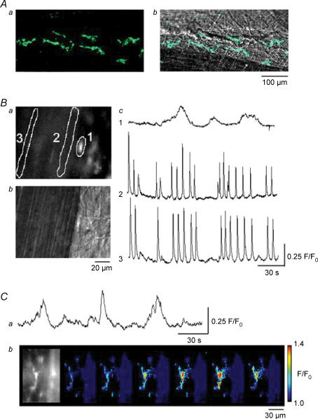
Figure 3. Ca2+ transients recorded from smooth muscles and interstitial cells in the guinea-pig bladder.
Kit-positive ICs having spindle-shaped cell bodies, some 80 μm in length and less than 10 μm in width, are shown located adjacent to a pair of muscle bundles (Aa). The same images were superimposed on the plane images of the smooth muscle bundles (Ab). In a preparation loaded with fluo-4, IC located near the muscle boundary had a higher fluorescence intensity than that of smooth muscle (Ba). A plane image with Nomarski optics visualized the cell body of IC (Bb). When Ca2+ transients were recorded from the IC (area 1) and from two smooth muscle areas (areas 2 and 3), synchronous Ca2+ waves were detected at areas 2 and 3 (Bc). However, IC generated slow Ca2+ transients independently from those of smooth muscles (Bc). In another fluo-4-loaded preparation which had been exposed to nifedipine (10 μm) for 30 min, IC continued to generate slow Ca2+ transients (Ca). A series of frames with intervals of 2 s demonstrates a Ca2+ transient originating from the IC (Cb).

