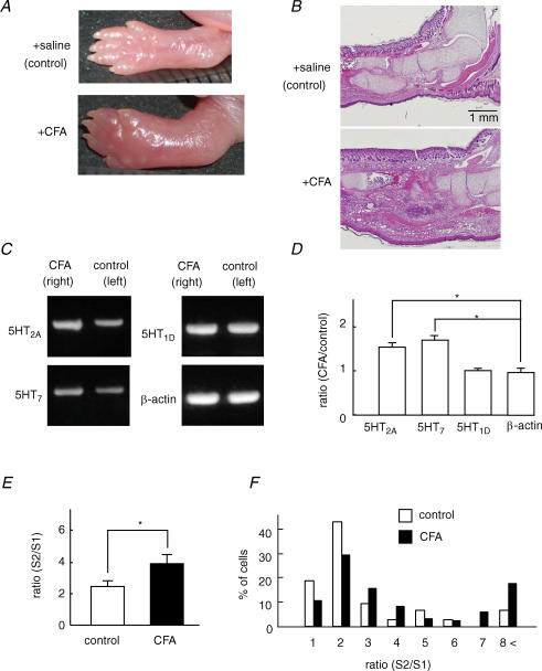
Figure 6. Inflammation increased 5-HT receptor expression and the potentiating effect of 5-HT on [Ca2+]i responses to capsaicin.
A and B, complete Freund's adjuvant (CFA) and saline were injected into the right and left hemilateral hind paw of the neonatal rat, respectively. Macroscopic (A) and histological (B) images 4 days after injection of CFA at the CFA-injected site (+CFA) and saline-injected site (control). C, the expression levels of 5-HT2A and 5-HT7 receptors were more pronounced in DRG isolated from the CFA side than the control side. No significant changes occurred in 5-HT1D and β-actin expression in DRG from both sides. D, the ratio of the optical density of PCR products of 5-HT2A, 5-HT7, 5-HT1D and β-actin from the CFA side to that from the control side (N = 5). * P < 0.05. E, the summarized potentiation rate (S2/S1) estimated by the ratio of the capsaicin (30 nm)-induced [Ca2+]i increase during the application of 5-HT (S2) to that before 5-HT application (S1). CFA, n = 114; control, n = 69; N = 5. * P < 0.05. F, histogram of the percentage of cells–S2/S1 ratio relation in response to capsaicin in DRG neurons isolated from the CFA side and control side.

