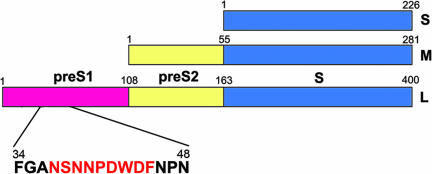
Fig. 1.
Domains of HBV surface proteins. The domain structures of S, M, and L proteins are presented with each domain colored differently. The adr subtype preS1 includes an epitope that is recognized by the neutralizing monoclonal antibody HzKR127. The peptide sequence of the epitope region is presented below the preS1 domain, with the epitope colored red.

