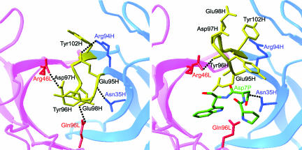
Fig. 3.
Comparison of antigen-binding site in the free and bound HzKR127 Fabs. Interactions between the H3 lid (yellow) and neighbors in the free (Left) and bound (Right) HzKR127 Fabs. The residues involved in the interactions are drawn, and the major interactions are presented as dotted lines. The light and heavy chains are colored pink and blue (labeled red and blue), respectively. The bound peptide residues Pro6P–Trp8P are presented as sticks with atomic colors.

