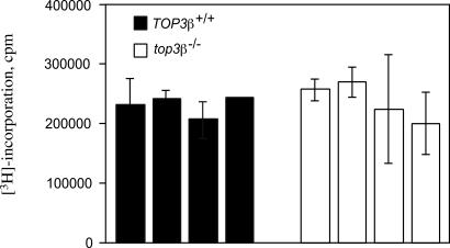
Fig. 3.
An example of T cell proliferation on stimulation with anti-CD3 antibodies. In this experiment, four 6-week-old TOP3β+/+ mice and the same number of their top3β−/− age-mates were used. Growth of T cells purified from the spleens of individual animals was stimulated by the addition of anti-CD3 antibodies to a final concentration of 0.1 μg/ml, and [3H]thymidine was added 48 h later to pulse-label replicating DNA. Data represent the average of triplicate cell samples from each of the four individual TOP3β+/+ and top3β−/− mice, and error bars represent standard deviations of the samples.

