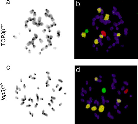
Fig. 6.
Aberrations in chromosome numbers in somatic top3β−/− cells. Two sets of bone marrow cell metaphases, one from a TOP3β+/+ and the other from a top3β−/− animal, are illustrated. Metaphase spreads were hybridized with a mixture of fluorescence-labeled probes to mark the autosomes 1, 3, and 16 in one color and the sex chromosomes in different colors; all chromosomes were further stained with DAPI before viewing in a fluorescence microscope. (a and c) The DAPI-stained chromosomes were more easily visualized as black-and-white images. In the examples shown, the TOP3β+/+ cell contained the full complement of 20 pairs of chromosomes, but one of the painted autosomes is missing in the top3β−/− bone marrow cell. (b and d) Separate fluorescent images of each set were merged to show the Cy5-labeled autosomes 1, 3, and 16 in pale yellow; the Cy3-labeled X chromosome in red; the FITC-labeled Y chromosome in green; and all of the other chromosomes in purple.

