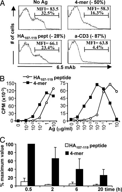
Fig. 1.
Influence of epitope multimerization on T cell responses in vitro and on half-life in vivo. (A) Effect on TCR down-regulation. 6.5 expression on gated CD4 cells from 6.5-TCR mice was analyzed by FACS after 12 h of stimulation with medium alone, monomeric HA107–119 peptide (10 μg/ml), 4-mer (10 μg/ml), or anti-CD3ε mAb (0.5 μg/ml). The decrease in 6.5+ CD4+ T cells compared with the unstimulated cells is shown in parentheses. (B) Effect on the proliferative response of HA-specific CD4 T cells (Left) Spleen and lymph nodes of 6.5-TCR mice were incubated with the indicated concentrations of the HA107–119 peptide or 4-mer, and [3H]thymidine incorporation was measured after 72 h of stimulation. Similar results were obtained with shorter (12, 24, and 48 h) stimulation periods (data not shown). (Right) TH1-differentiated 6.5-TCR CD4 T cells were incubated for 24 h with the indicated concentrations of the HA107–119 peptide or 4-mer and irradiated APCs, and [3H]thymidine uptake was evaluated. Results are from one of two similar experiments. (C) In vivo half-life on splenic APCs of the HA107–119 peptide or 4-mer. Splenocytes from BALB/c mice, injected i.v. with 2.5 μg of the HA107–119 peptide or 4-mer, were harvested at the indicated time points to induce proliferation of purified 6.5-TCR CD4 T cells. Maximum [3H]thymidine uptake (62,885 ± 11,655 cpm) was defined as the stimulation induced by splenocytes from BALB/c mice injected 0.5 h earlier with 4-mer. Results from three experiments are presented as the mean percentage (± SEM) of the maximum value.

