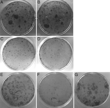
Fig. 2.
Evaluation of KSC cfus in VDR-null mice. Keratinocytes isolated from VDR-null mice (B, D, and F), wild-type mice (A, C, and E), and a VDR-null mouse expressing a keratinocyte-specific VDR transgene (G) were plated onto irradiated 3T3 cells and cultured for 2 (C and D) or 4 (A, B, and E–G) weeks. Cells were isolated from 3-day-old (A and B) and 4-week-old (C–G) mice. Staining was performed with rhodanile blue. Data are representative of those obtained with at least three different mice of each age and genotype.

