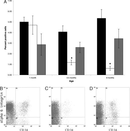
Fig. 4.
Quantitation of CD34/α-6 integrin-positive KSCs. (A) Keratinocytes were isolated from wild-type (black bar) and VDR knockout littermates lacking (white bar) and expressing (gray bar) a keratinocyte-specific VDR transgene at 1, 3.5, and 9 months of age. Cells were subjected to FACS analysis to determine the number of doubly labeled cells. Numbers represent the mean ± SEM of cells isolated from at least three different mice of each genotype. ∗, P < 0.05. A representative cell-sorting profile from 9-month-old wild-type (B), VDR-null (C), and keratinocyte-specific VDR transgene-positive, VDR-null (D) mice is presented.

