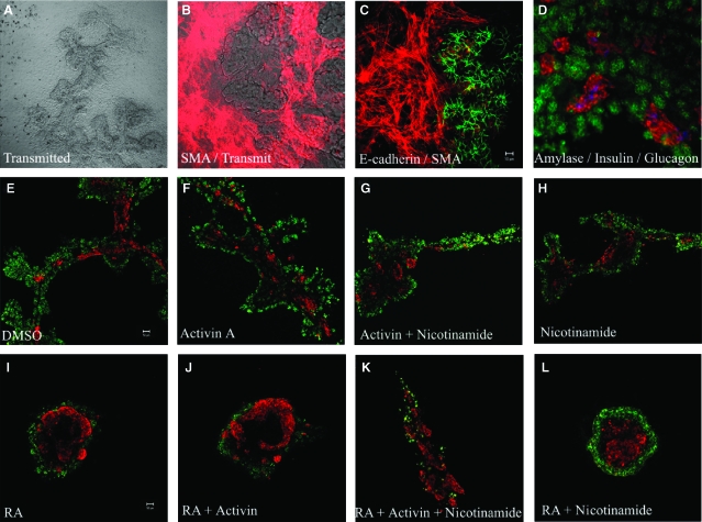
Fig. 1.
Differentiation of dorsal pancreatic buds in vitro. (A) Seven-day cultured pancreatic bud, the epithelium forms an extended branched structure. (B, C) The cultured pancreatic buds flatten out onto the substratum and mesenchymal cells spread rapidly out of the explant to form a monolayer of cells which stain for smooth muscle actin (red). The pancreatic epithelium was stained positive for E-cadherin (green). (D) Buds were cultured for 7 days, fixed, and stained for amylase (green), insulin (red), and glucagon (blue). Pancreatic buds were cultured for 2 days and then treated for 5 days with (E) 0.1% DMSO, (F) 10 μg/ml activin A, (G) 10 μg/ml activin A and 5 mM nicotinamide, (H) 5 mM nicotinamide (I) 1 μM all-trans retinoic acid (atRA), (J) 1 μM atRA and 10 μg/ml activin A, (K) 1 μM atRA, 10 μg/ml activin A, and 5 mM nicotinamide (L) 1 μM atRA and 5 mM nicotinamide. Buds were fixed and stained for amylase (green) and insulin (red).

