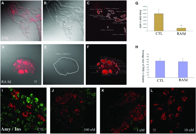
Fig. 2.
Effect of all-trans retinoic acid (atRA) on differentiation and branching morphogenesis of pancreatic buds in culture. Two-day pancreatic buds were cultured for 5 days without (A–C) or with 1 μM retinoic acid (RA) (D–F) and stained for insulin (red). (A) Overlay image of transmitted light image (B) and fluorescence image (C), (D) overlay image of transmitted light image (E) and fluorescence image (F). Area analysis in (A–C) and (D–F) was performed by Zeiss image processing software LSM 5. (G) Area of control and RA-treated pancreas (in μm2). The total areas were calculated from three bud cultures. (H) The area of insulin-expressing clusters was measured by LSM software. Large clusters (>500 μm2) were calculated from three buds and the averaged results are shown as means+SD. (I–L) 2-day pancreatic buds were cultured for 5 days with (I) 0.1% DMSO, (J) 100 nM, (K) 1 μM, (L) 10 μM atRAs and stained for amylase (green) and insulin (red).

