Fig. 5.
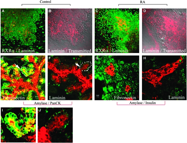
Up-regulation of extracellular laminin by retinoic acid. E11.5d dorsal pancreatic buds were isolated, culture for 2 days and then cultured for 3 days (A–D), or 5 days (E–H) without (A, B, E–H) or with 1 μM retinoic acid (C, D). Buds were then fixed and stained for (A, C) retinoid-X receptorα (RXRα) (green)/laminin (red). (B, D) Overlay picture of laminin staining with transmitted images of (A) and (C). (E, F) Buds were stained for amylase (green)/Pan-cytokeratin (red). (G, H) Buds were stained for amylase (green)/insulin (red). In (F, H) pancreatic buds were cultured on laminin-coated coverslips for 7 days. Laminin mimics the effect of all-trans retinoic acid on exocrine differentiation. I and J are high-powered images from E and F.

