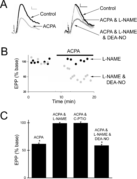
Fig. 5.
Arachidonylcyclopropylamide (ACPA)-induced synaptic inhibition requires nitric oxide. (A) Representative end-plate potentials (EPPs) recorded before and after the application of ACPA (10 µm). Either Nω-nitro-l-arginine methyl ester (L-NAME) or L-NAME and diethylamine/NO complex (DEA-NO) (traces on the right) were present during the recording of the EPPs. Each trace represents the average of eight sweeps. Resting membrane potentials were approximately −90 mV. Calibration bars, 0.5 mV, 2 ms. (B) Time course of EPP amplitudes from two representative experiments. The application of ACPA (10 µm) to a neuromuscular junction (NMJ) exposed to either L-NAME (black) or L-NAME and DEA-NO (grey) is indicated by the horizontal bar. Each data point represents the amplitude of an average of eight AC-coupled sweeps. (C) Mean percent reduction of EPP amplitudes (from initial baseline readings) following 5–10 min of ACPA (10 µm) application. ACPA was applied either alone (n = 11), with L-NAME (0.3 mm, n = 5), with 2-(4-carboxyphenyl)-4,4,5,5-tetramethylimidazoline-1-oxyl-3-oxide potassium salt (C-PTIO) (40 µm, n = 4) or with L-NAME and DEA-NO (0.1 mm, n = 5). *The mean EPP amplitude is significantly different from when it was measured under baseline conditions (P < 0.05; Student's t-test). Baseline measurements were made either under control conditions, in the presence of L-NAME, in the presence of C-PTIO or in the presence of L-NAME and DEA-NO, as appropriate for the experiment. Error bars represent SEM.

