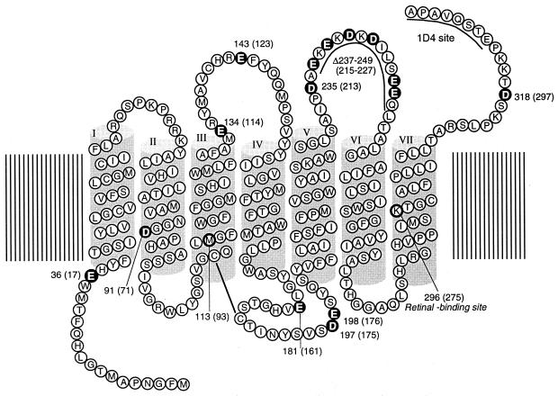
Figure 1.
Secondary structural model of retinochrome showing the location of Glu and Asp residues (white letters). For affinity purification with an anti-rhodopsin antibody, the amino acid sequence of monoclonal antibody Rho1D4 epitope (ETSQVAPA) is introduced to the C terminus of retinochrome. On the basis of sequence alignment, the bovine rhodopsin amino acid residue numbering system is used. The retinochrome numbering system is shown in parentheses.

