
Figure 7.
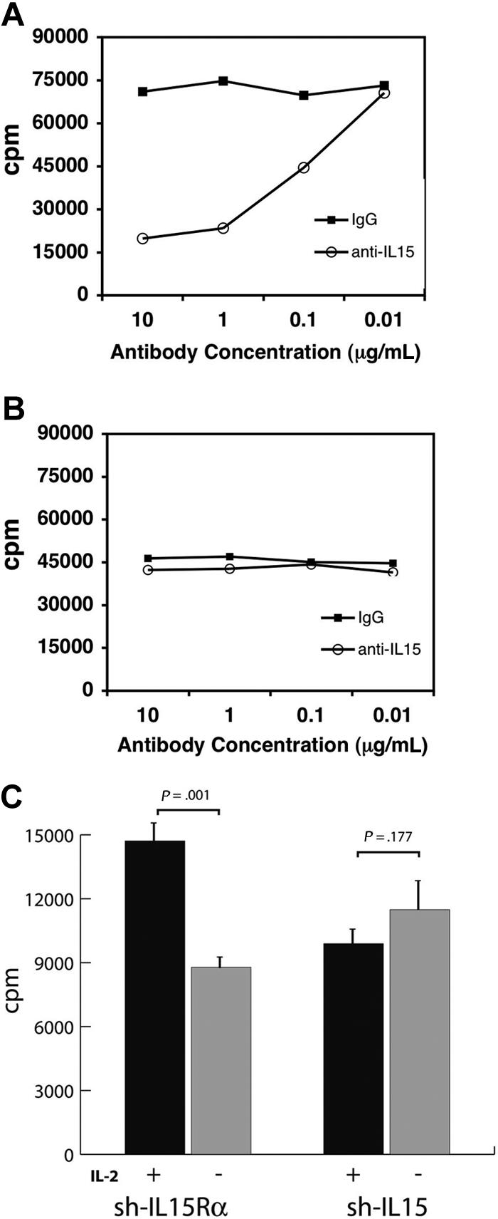
LC15 proliferation is not affected by α–IL-15 blocking antibody. For each assay, 1 × 10⁵ cells were plated in triplicate in 96-well plates. Cells were cultured for a total of 24 hours. [3H]-thymidine was added to cultures 18 hours prior to scintillation counting. (A) To confirm activity of the α–IL-15 antibody, normal, activated T cells were plated with indicated concentrations of either α–IL-15 antibody or matched isotype control antibody. Subsequently, 10 ng IL-15/mL was added to the culture media. (B) To evaluate the effect of α–IL-15 antibody on LC15 cells, LC15 cells were washed and plated with serial dilutions of either α–IL-15 antibody or matched isotype control antibody. No inhibition of LC15 cells was seen. (C) Retroviral vectors containing shRNAs directed to either the human IL-15Rα or endogenously expressed human IL-15 were used to transduce LC15 cells and cells cultured in medium containing IL-2 and puromycin. Selected cells were withdrawn from IL-2–containing medium for 3 days, and their proliferation was measured by [3H] thymidine incorporation. [3H]-thymidine was added to cultures 48 hours prior to scintillation counting. Standard deviations indicated by error bars. Significance was evaluated by Student t test.