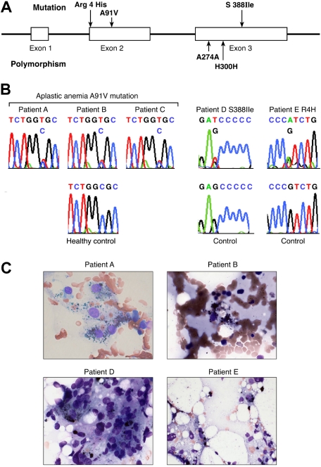
Figure 1.
Perforin mutations in patients with aplastic anemia. (A) Linear structure of the PRF1 gene, which encodes the human perforin protein and aplastic anemia-associated mutations. The segments represent the exons. PRF1 has 3 exons, 2 of which (exons 2 and 3) contain coding sequences. In aplastic anemia, nonsynonymous mutations were found in exon 2 (codon 4 Arg/His and codon 91 Ala/Val) and exon 3 (codon 388 Ser/Ile). The polymorphisms in exon 3 are also shown (codons 274 and 300). R indicate arginine; H, histidine; I, isoleucine; V, valine; A, alanine. (B) Sequences of the patients carrying the mutations compared with sequences obtained from the controls. Green identifies adenine (A); red identifies thymidine (T); black identifies guanine (G); and blue identifies cytosine (C). (C) Bone marrow smear examination from the patients carrying PRF1 mutations. Four of 5 patients carrying mutations revealed hemophagocytosis in bone marrow at diagnosis.

