Fig. 1.
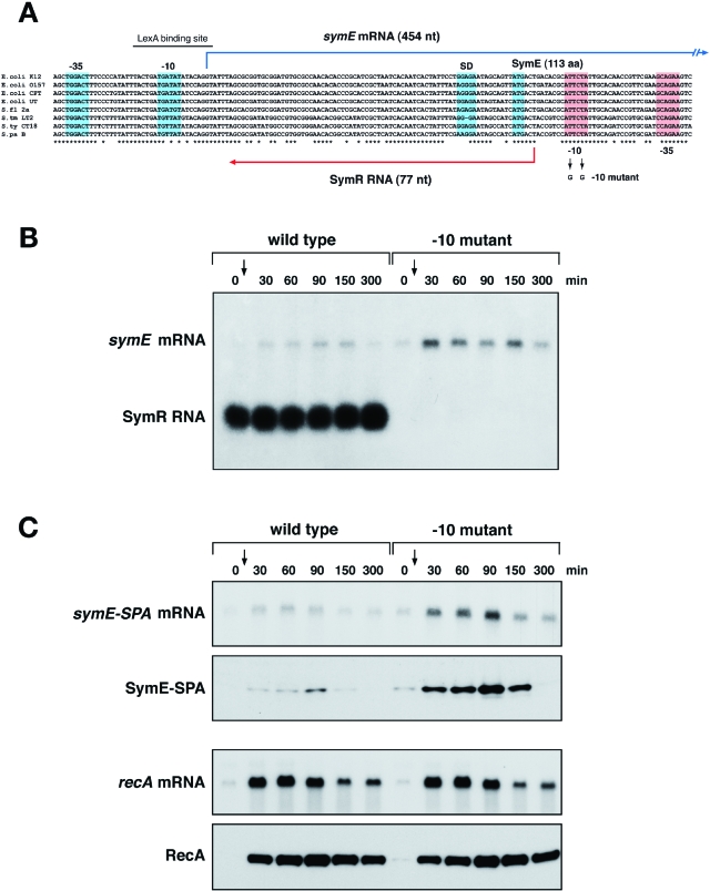
SymR RNA represses SymE translation. A. Genetic organization of the symER locus. Multiple sequence alignments of the different symER sequences were constructed using clustalw. The species correspond to E. coli strains K12, O157:H7, CFT073 and UT189, Shigella flexneri 2a, Salmonella typhimurium LT2, S. typhi CT18 and S. paratyphi B. The −10 and −35 promoter sequences, the Shine-Dalgarno sequence and initiation codon of symE are indicated by blue boxes. The −10 and −35 sequences of symR are indicated by red boxes. B. symE mRNA and SymR RNA levels in MG1655 and the −10 symR promoter mutant. Total RNA was isolated from wild-type MG1655 and −10 mutant strains grown in LB medium at 37°C at 0, 30, 60, 90, 150 and 300 min after treatment with 1 μg ml−1 mitomycin C. Samples (5 μg) were analysed by Northern hybridization using oligonucleotide probes specific to symE and SymR. C. symE-SPA mRNA and protein and recA mRNA and protein levels in MG1655 symE-SPA and the −10 symR promoter mutant. Total RNA and cell lysates were prepared from MG1655 symE-SPA and symE-SPA −10 mutant strains in grown in LB medium at 37°C at 0, 30, 60, 90, 150 and 300 min after treatment with 1 μg ml−1 mitomycin C. RNA samples (5 μg) were analysed by Northern hybridization using oligonucleotide probes specific to symE and recA, and cell lysates were analysed by immunoblot assays using monoclonal anti-FLAG M2-AP and polyclonal anti-RecA antibodies.

