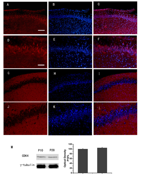
Figure 1.

Expression and localization of cyclinD1-CDK4 during the postnatal development in hippocampus area CA1. Immunofluorescent microscopic localization of CDK4 in hippocampus area CA1 at PND10 (A-F) and at PND28 (G-L). Blue indicates nuclear DAPI counterstain, and red indicates CDK4. C, F, I, L are the merged image. Scale bar: in A (applies to A-C and G-I), 121 μm; in D (applies to D-F and J-L), 48 μm. Detection of CDK4 on Western blots (M). PND (postnatal day)
