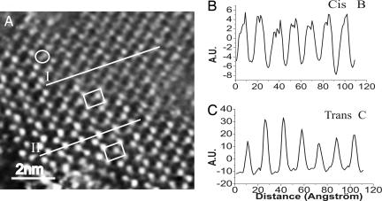
Fig. 4.
Cooperative switching occurring over a single molecular domain. (A) STM image of SAM of 1 formed on stripped Au(111) incubated in the dark for one night in 5·10−4 M solution in toluene. In the image, a single cis molecule is marked by the oval. The sample was irradiated for 15 min before measurements. The image was recorded 90 min after the irradiation. This time frame is very small if compared with the 2 weeks needed to achieve a complete restoration of the trans isomer of alkyl-thiolated cis-azobenzenes under dark (42). The lower unit cell contains the trans isomers; the upper unit cell contains cis molecules. The profiles are traced along the direction of the unit cell main axis, i.e., the direction of the switch (see Fig. 2B) IT = 49.00 pA; VT = 145 mV. (B and C) Line profiles showing profile I the cis conformer (B) and profile II the trans conformer (C). The two spots were observed to be located in identical positions also upon changing systematically the scan angle and scan rate, as well as on different samples and employing different tips, therefore ruling out any imaging artifact. Moreover, the switch was visualized by STM by using various tunneling parameters and therefore diverse tunneling gap impedances. Thus, although we cannot fully neglect a mechanical or electrical contribution of the tip to the switch, we do believe it does not hold a prime role. Although the isomerization occurs over some hundreds of adjacent molecules, it does not take place over the whole sample surface.

