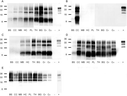
Figure 5. PrPres Distribution Profiles in Brains of Atypical and Classical Scrapie–Affected SRs by WB.
Atypical scrapie cases S5/FS (A), S6/FS (B), and S7/CS (D) in sheep and G2/FS (C) in a goat compared to a classical scrapie reference case from Switzerland (E). For comparison between individual membranes, the same amount of a classical scrapie sample (+) was loaded onto each gel. Neuroanatomical structures: BS, caudal brainstem; CC, cerebellar cortex; MB, midbrain; HC, hippocampus; PL, piriform lobe; TH, thalamus; BG, basal ganglia; CF, frontal cortex; CO, occipital cortex. TSE-negative brain tissue served as negative control (−).

