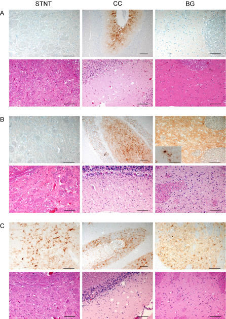
Figure 6. IHC and Histopathology of Selected Brain Structures in Atypical and Classical Scrapie–Affected SRs.
Comparison of PrPsc immunolabeling with monoclonal antibody F99/97.6.1 in IHC (upper panel) and vacuolar lesion in histopathology (lower panel) in atypical scrapie cases S6/FS (A) and S7/CS (B) in sheep, compared to a classical scrapie reference case from Switzerland (C) in different neuroanatomical brain structures: brainstem, spinal tract nucleus of the trigeminal nerve (STNT); cerebellar cortex (CC), and basal ganglia (BG).
(B) The inset for S7/CS, basal ganglia, shows plaque-like PrPsc deposits in the ventral and the medial geniculate body of the thalamus. Bars indicate 100 μm, except for the IHC of CC (150 μm).

